Pekkan Lab
Research
Connecting biofluid mechanics, cardiovascular biology, imaging, and engineering to understand disease and develop new approaches to treatment.
Research area · 01
Computational blood flow dynamics
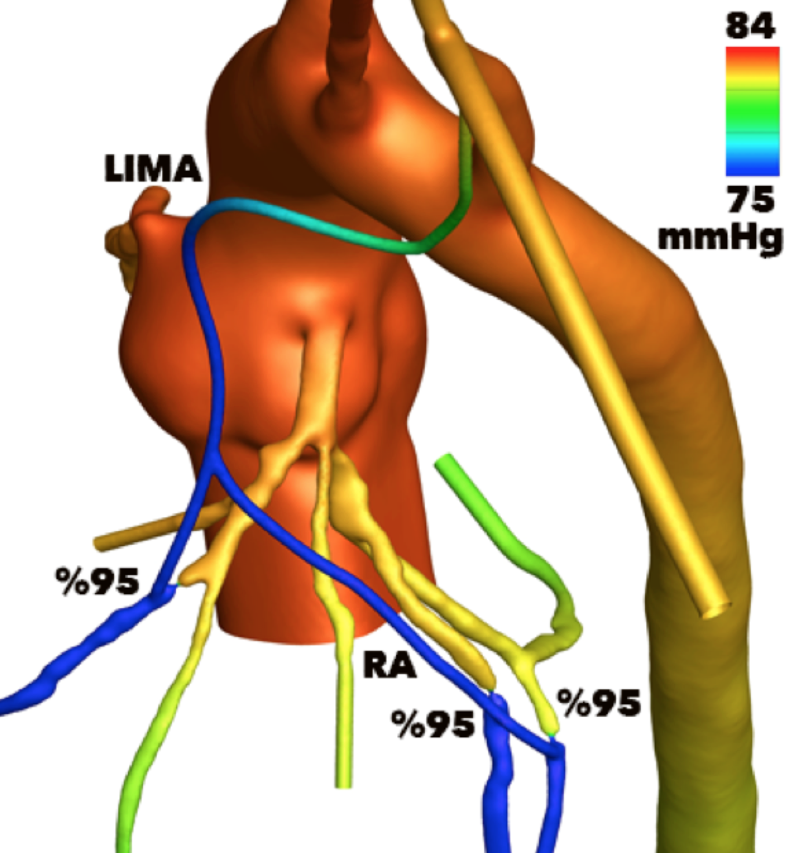
We use computational fluid dynamics (CFD) to model blood flow in the cardiovascular system in health and disease. These models help us investigate complex flow patterns, estimate hemodynamic quantities that are difficult to measure directly, and assess how the circulation may respond to clinical interventions. Our goal is to support diagnostic assessment and the evaluation of treatment options.
Research area · 02
Patient-specific surgical planning
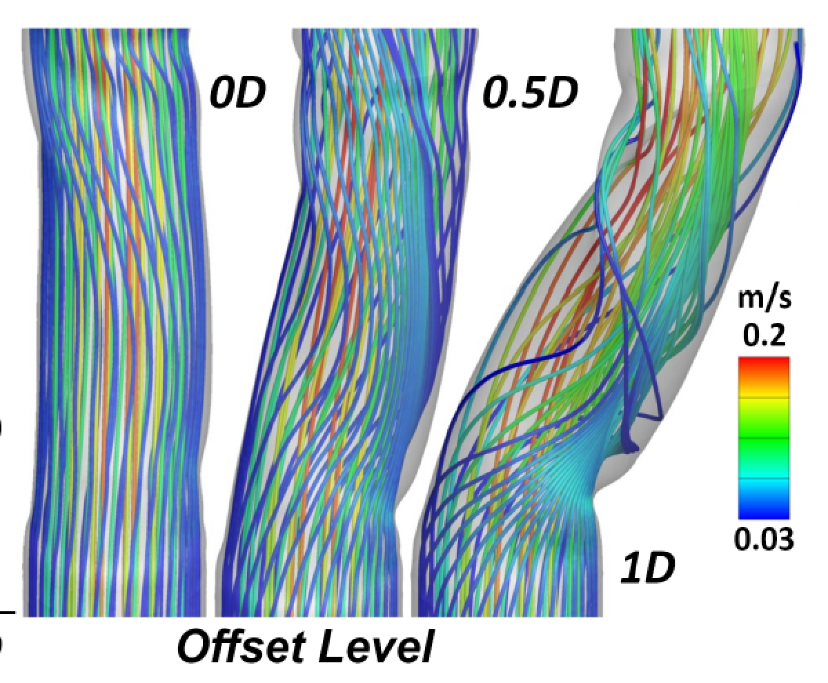
We develop patient-specific computational models to support planning for pediatric cardiovascular surgery. Our methods compare the predicted blood flow and mechanical performance of alternative surgical configurations before an intervention. This work includes patented approaches to cardiovascular modeling and surgical planning, with the aim of helping clinicians evaluate options for each patient’s anatomy and physiology.
Research area · 03
Imaging cardiovascular development
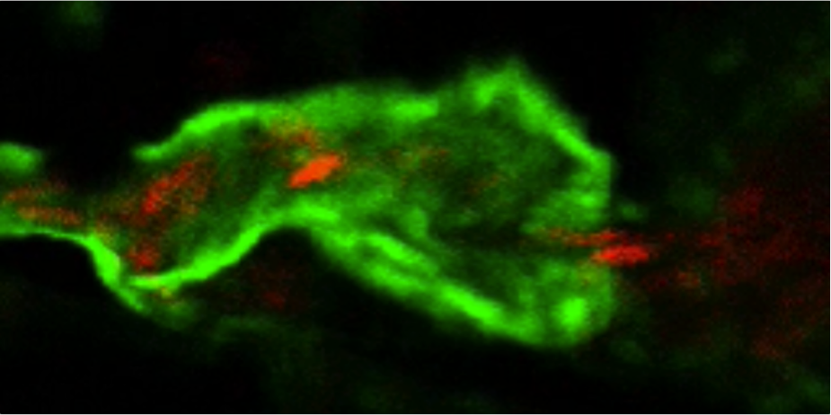
We use imaging of genetically engineered zebrafish and mouse models to study cardiovascular development and interactions between the cardiovascular and nervous systems. By visualizing vessels and blood cells in three dimensions, we investigate how biological structures and blood flow develop together. This work aims to improve our understanding of mechanisms relevant to cardiovascular disease.
Research area · 04
Soft tissue engineering
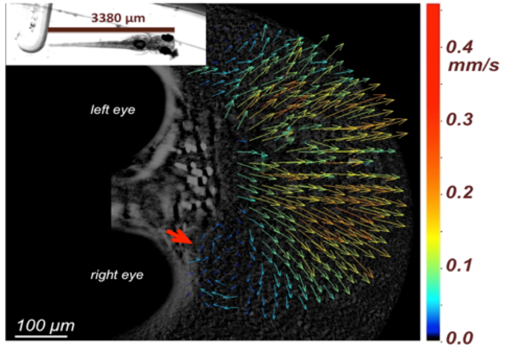
We investigate how mechanical forces influence the development and remodeling of cardiovascular tissues, with a focus on congenital heart defects. Our studies examine relationships between blood flow, vascular growth, and gene expression in the right vitelline artery and aortic arches of chick embryos [1], [2]. We combine changes in mechanical loading and genetic conditions with quantitative imaging to investigate how these factors affect tissue development and disease progression.
Research area · 05
Cardiovascular biology
We study the structure and surface characteristics of blood vessels used in cardiovascular surgery, including the left and right internal mammary arteries, great saphenous vein, and carotid artery. In a study involving 54 patients undergoing off-pump coronary artery bypass surgery, we used scanning electron microscopy (SEM) to compare arterial and venous graft samples across graft types and patient risk groups. We also investigated the effects of two graft-preparation solution concentrations on thrombogenic characteristics. Transmission electron microscopy (TEM) provides a complementary approach for examining intracellular structures in these tissues.
Research area · 06
Cardiovascular devices
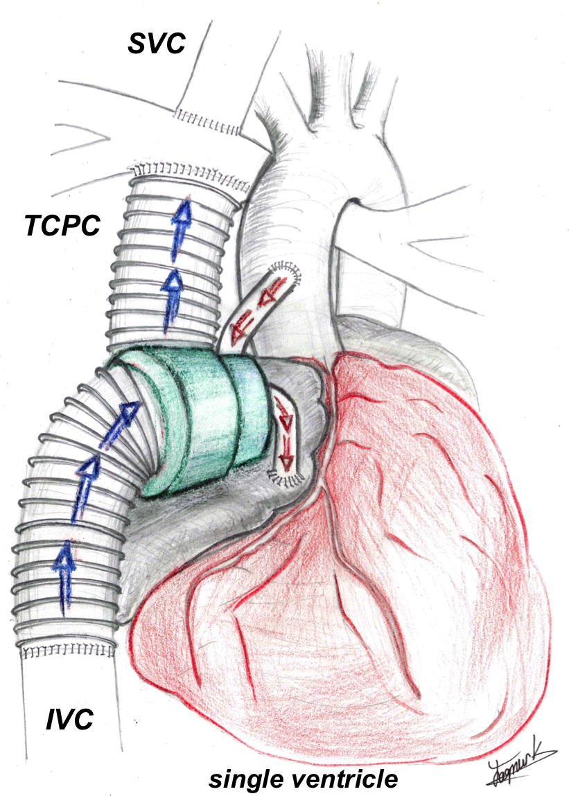
We develop and evaluate cardiovascular devices, with a particular focus on circulatory support for patients with single-ventricle congenital heart disease. Our integrated aortic-turbine venous-assist device (iATVA) couples an aortic turbine to a venous pump and has been designed and tested in vitro as a potential approach to support the Fontan circulation [3]. We use CFD to investigate and optimize device performance. Our research also includes polymeric artificial heart valves for right ventricular outflow tract (RVOT) reconstruction, alongside the development of manufacturing and implantation methods.
Our facilities
Equipment & capabilities
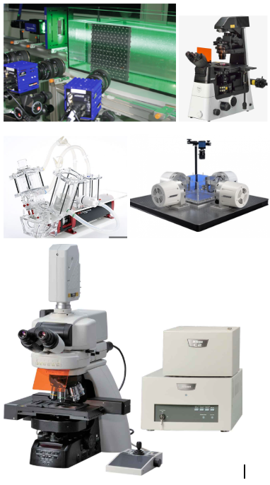
- Tomographic particle image velocimetry (PIV) system with three Phantom M310 high-speed cameras, one standard camera, two LaVision lasers, optics, safety equipment, and acquisition hardware and software.
- Biaxial mechanical testing system with two 200 N and two 100 N actuators, originally supplied by Bose ElectroForce.
- ViVitro pulse duplicator with an organ-culture circuit separator, impedance adapter, and data-acquisition computer; two in vitro Transonic flowmeters with probes; one in vivo Transonic flowmeter with microartery probes; in vivo and in vitro pressure measurement systems; and an electromagnetic flowmeter.
- Nikon C2si confocal microscope with an Okolab environmental chamber for live-cell imaging, a custom egg-incubation chamber, three objectives, and four lasers.
- Two servo-null pressure measurement systems for embryonic heart studies.
- Leica MZ16 FA microscope with temperature-controlled plate, filters, and associated accessories.
- Narishige glass-pipette preparation equipment, including the MF-900 Micro Forge, a puller, and a polisher.
- Nikon Eclipse Ti2-U inverted microscope for tissue-culture imaging, with objectives and cameras.
- Thorlabs three-dimensional probe-based optical coherence tomography (OCT) system.
- Tissue preparation and immunohistochemistry equipment, including a Leica RM2125 RTS microtome, Leica EG1150 C cold plate, fume hood, and TEM crystals.
- Formlabs Form 2 stereolithography (SLA) printer.
- 3D Systems ProJet 660 Pro powder-based printer.
References
- Goktas S, Uslu FE, Kowalski WJ, Ermek E, Keller BB, Pekkan K. Time-Series Interactions of Gene Expression, Vascular Growth and Hemodynamics during Early Embryonic Arterial Development. PLOS ONE, 11(8), Article e0161611 (2016). DOI: 10.1371/journal.pone.0161611.
- Karakaya C, Goktas S, Celik M, Kowalski WJ, Keller BB, Pekkan K. Asymmetry in Mechanosensitive Gene Expression during Aortic Arch Morphogenesis. Scientific Reports, 8(1), Article 16948 (2018). DOI: 10.1038/s41598-018-35127-7.
- Pekkan K, Aka IB, Tutsak E, Ermek E, Balim H, Lazoglu I, Turkoz R. In vitro validation of a self-driving aortic-turbine venous-assist device for Fontan patients. The Journal of Thoracic and Cardiovascular Surgery, 156(1), pp. 292–301.e7 (2018). DOI: 10.1016/j.jtcvs.2018.02.088.